|
Case of the Month: December 2025
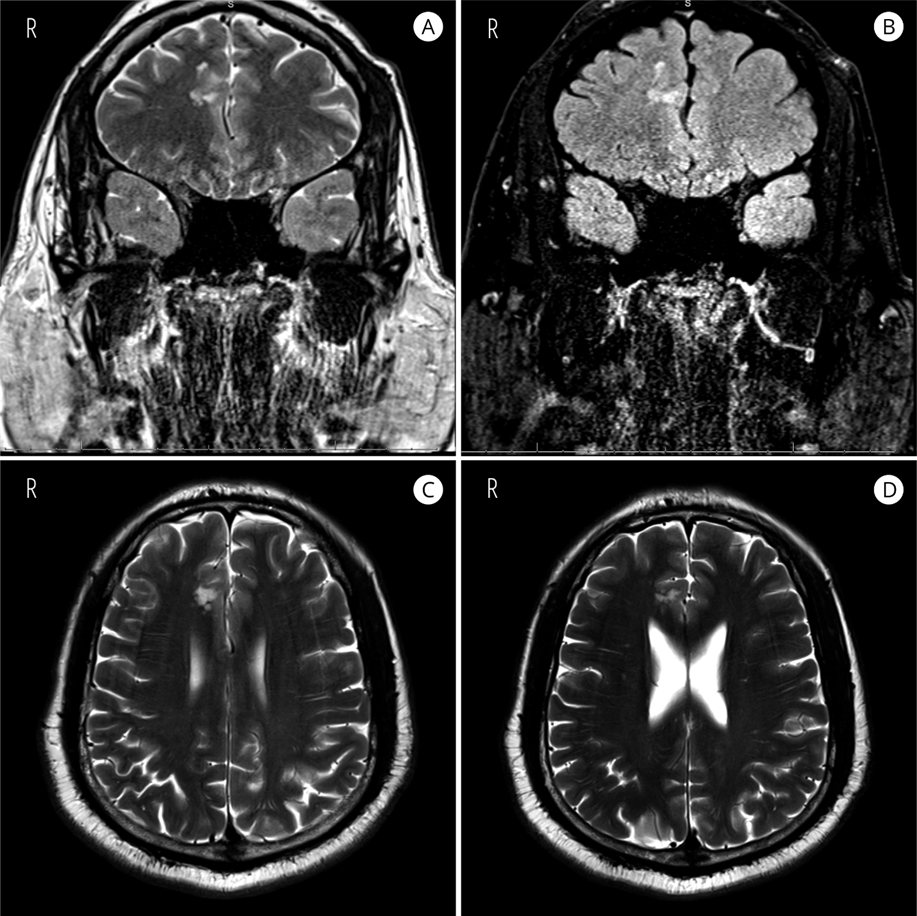
Title: A 53-year-old male with a history of intractable epilepsy. Author: Powell, Reuben P., Tursunbaev, Farkhod, and Campbell, Gerald A. Institution: The University of Texas Medical Branch Clinical History: A 53-year-old male with a history of intractable epilepsy since adolescence presented for evaluation. MRI revealed nodular, non-enhancing subcortical T2/FLAIR hyperintense lesions in the right parasagittal frontal lobe, along the cingulate gyrus and sulcus, measuring 18 x 13.2 x 12.8 mm in aggregate. The patient underwent complete surgical resection and became seizure-free postoperatively with tapering of antiepileptic medications. Radiology:
Representative Histology/IHC:
 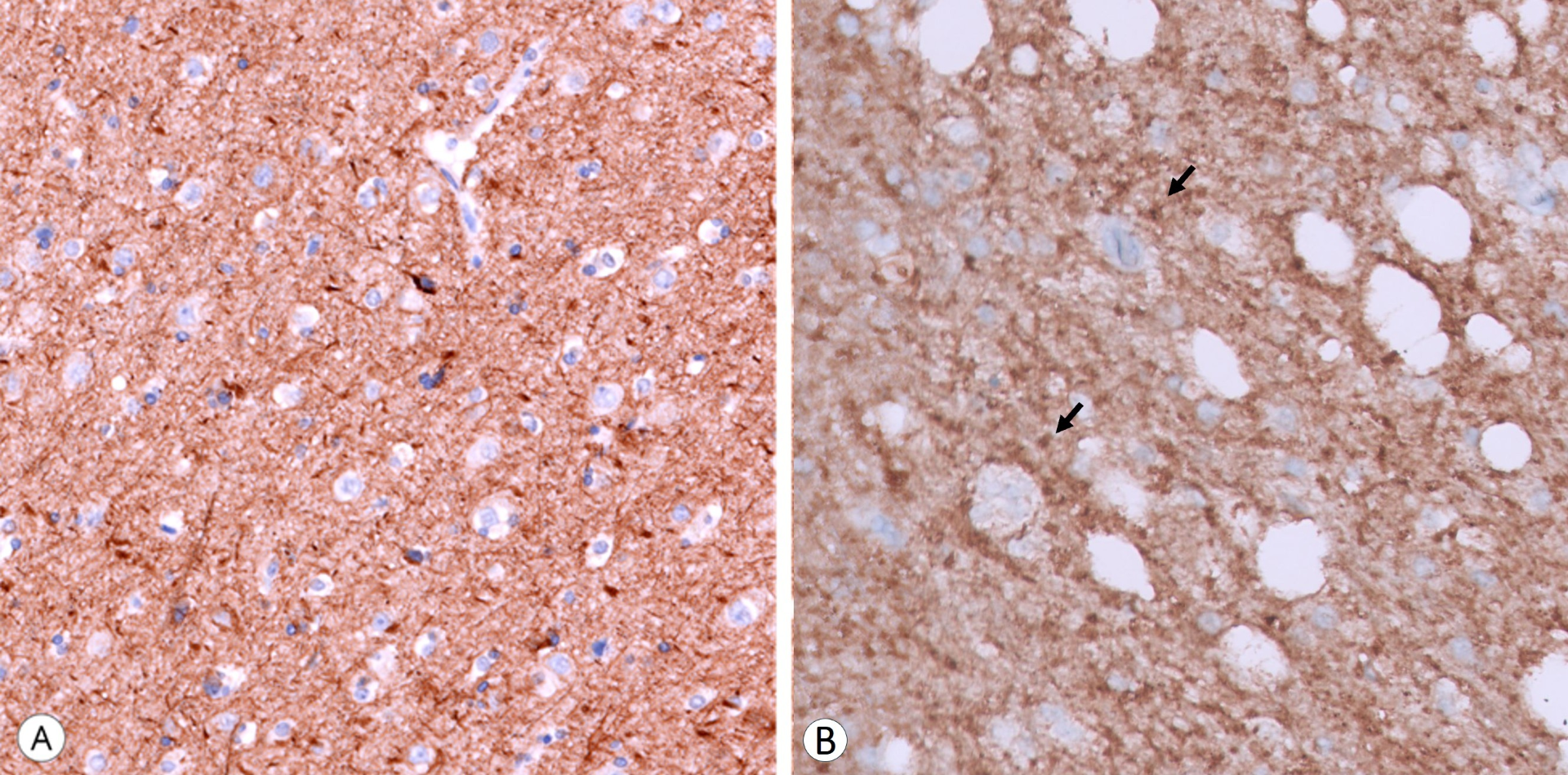
Questions for Viewers: What is the differential diagnosis for these radiologic and histologic findings? How does immunoreactivity for OLIG2 inform the distinction among low-grade epilepsy-associated tumors, given the overlapping expression of OLIG2 in glial and some glioneuronal neoplasms? |
Our Sponsors