
|
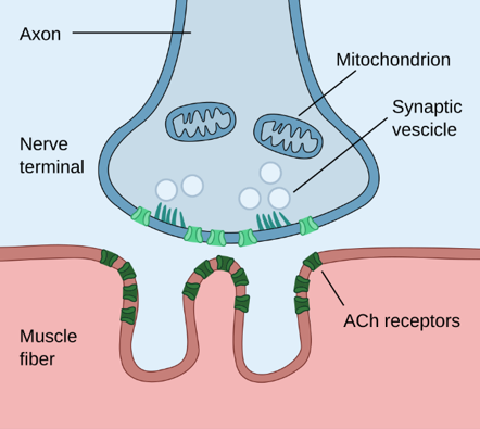
Diagnosis and Discussion The electron micrograph shows a normal neuromuscular junction (synapse) and has no diagnostic significance. At the neuromuscular junction, the innervating axon is adjacent to the motor end plate, which has a corrugation of the muscle fiber’s sarcolemmal surface (see diagram below).
Diagram of a neuromuscular junction, showing acetyl choline receptors and mitochondria in the nerve terminal, synaptic cleft, and folded membrane of the motor endplate of the myocyte. Author: Paul Hege, own work, CC by SA 4.0; Date: 22 March 2020; Published on Wikipedia under image sharing policies, “Neuromuscular Junction”. By Paul Hege - Own work, CC BY-SA 4.0, https://commons.wikimedia.org/w/index.php?curid=88461302
The axon terminal (axonal bouton) is filled with synaptic vesicles, and correspondingly in the electron micrograph, the vesicles are indeed packed in the bouton and within the synaptic cleft. Although the light microscopic findings of rounded, variably-sized myocytes and interstitial connective tissue would be quite abnormal for a quadriceps biopsy, this is the normal histologic appearance of extraocular muscles. The diagnosis is based on light microscopy and notes the pertinent negatives for the clinical differential. Most would report the findings as below: Medial Rectus Biopsy with Type 2 fiber predominance. No evidence of amyloid deposition. No evidence of mitochondrial structural abnormalities. Discussion: This case and questions highlight the importance of understanding the physiologic variability between muscle groups, particularly the variability in myocyte size. The questions also highlight the strategies of writing up a negative report to include pertinent negatives of diagnostic differentials to adequately communicate the pathologic findings to the treating clinician. Below the differentials are discussed with the expected clinical scenarios and histologic findings. Differentials: The differentials in this case included amyloidosis, chronic progressive external ophthalmoplegia, and other mitochondrial disorders. Amyloidosis: Amyloid deposition can occur in the extraocular muscles with horizontal recti, including medial and lateral rectus muscles, considered the most common. Clinical: Symptoms most commonly include diplopia, ophthalmoplegia, and proptosis. Patients with systemic amyloidosis are more likely to have multiple extraocular muscles involved bilaterally, as well as other organ systems and thus additional symptoms. Radiology: MRI is considered an important diagnostic tool; however, there can be variable findings with the most common being: fusiform shaped enlargement, intralesional heterogeneity, and contrast-enhancement. Pathology: Muscle biopsy is necessary to confirm extra-ocular muscle (EOM) amyloidosis, with appearance of homogenous eosinophilic extracellular material on H&E and positive Congo red staining with apple green birefringence under polarized light. Subtyping of amyloid, by mass spectroscopy or immunohistochemistry, is recommended for determination of best treatment for EOM amyloidosis as management varies from debulking, radiation, and conservative monitoring. Mitochondrial disorders including Chronic Progressive External Ophthalmoplegia (CPOE): Mitochondrial disorders, specifically single large-scale mitochondrial DNA deletion syndromes (SLSMDS) including CPOE are more frequently diagnosed in younger populations, often before 20 years of age. Though this patient was elderly, the symptomology is fitting and thus these disorders should still be ruled out when there is clinical suspicion. The 2 most fitting SLSMDS’s for this patient would be CPOE and Kearns-Sayre syndrome (KSS). Clinical: Presenting symptoms for CPOE can include ptosis, ophthalmoplegia, proximal limb myopathy, dysphagia, and exercise intolerance. KSS presenting symptoms can include all the symptoms for CPOE as well as pigmentary retinopathy, which is the distinguishing feature between the two mitochondrial disorders. KSS can also include cardiac conduction abnormality. Radiology: MRI would demonstrate characteristic hyperintense spongiform signal in the recti muscles. Pathology: The pathophysiology and thus histology of CPOE and KSS are nearly identical, and therefore the delineation of either disease is more of a clinical and gene specific distinction. Most cases are associated with single large mitochondrial DNA (mtDNA) deletions; however, CPOE can be inherited as multiple small mitochondrial DNA deletions in autosomal dominant or autosomal recessive patterns. The deletions of the mtDNA creates a proliferation of the mtDNA. This increase in mtDNA can be seen histologically as subsarcolemmal accumulation of ragged red fibers on Gomori trichrome stain or increased expression of succinate dehydrogenase (SDH) as ragged blue fibers. When muscle fibers overexpress SDH, there is subsequently deficient staining for cytochrome oxidase (COX) as cox-negative fibers. See photo below (McClelland et al. 2016). Diagnostic tests include molecular genetic testing of the mitochondrial genome and compressive genomic testing; which can be taken from the leukocyte DNA in blood from children. Currently skeletal muscle biopsy is still recommended in adults to confirm the diagnosis.
Left: Cytochrome oxidase stain showing pale, COX-negative fibers (arrows). Center: Succinate dehydrogenase (SDH) stain showing the same fibers as overreactive “ragged-blue” (arrows). Right: Gomori trichrome stain showing a Bragged-red^ fiber. (MRI images courtesy of Stacy Pineles, MD, Jules Stein Eye Institute, University of California Los Angeles. Muscle pathology images courtesy of Alan Pestronk, MD, Washington University in St. Louis): Reference 4, below. References
|

Our Sponsors