
|
AANP New Case of the Month: September 2026
Title: Muscle biopsy for evaluation of progressive diplopia and external ophthalmoplegia
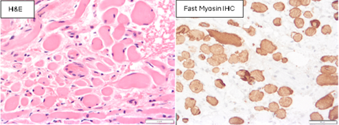
Authors: Drs. Danielle Lombardo, Doug Anthony, and Mike Punsoni Institution: Brown University Clinical History: A 76-year-old man had been followed by his ophthalmologist for worsening diplopia, exotropia, ptosis, and retinitis pigmentosa. The differential diagnosis was chronic progressive external ophthalmoplegia (CPEO), other mitochondrial disorder, or amyloidosis. During orbital surgery for strabismus (to reduce his diplopia) a biopsy of the medial rectus was performed to assess mitochondrial structure and to evaluate the possibility of amyloid deposition. Biopsy Findings The biopsy showed the histologic appearance of extraocular skeletal muscle, with rounded fibers, fiber size variability, and some intervening collagen. The majority of fibers expressed fast myosin. No abnormalities were detected on muscle biopsy histochemical special stains, and Congo Red staining was negative for amyloid.
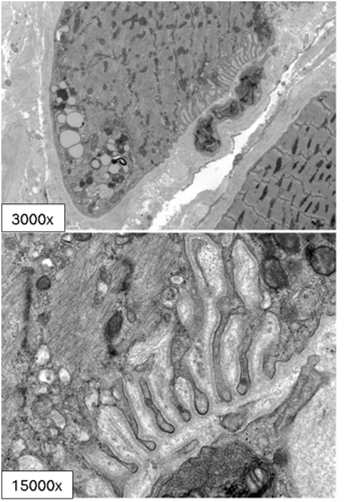
Electron microscopy was performed to assess mitochondria and showed normal ultrastructure of mitochondria (Direct mag 3000x and 15000x, respectively). However, there was an unusual, folded membrane alteration with aggregates of mitochondria in one myocyte (Electron micrographs below).
Questions for Participants: 1) What is the diagnostic significance of this EM finding? 2) How should the findings on this muscle biopsy be reported?
|
Our Sponsors